PotM: Disulfide-Based Self-Immolative Linkers

PotM: Disulfide-Based Self-Immolative Linkers

Published on 26/01/2021

The linkage of a drug to its carrier via a disulfide-based self-immolative linker allows for the specific intracellular release of the active molecule upon glutathione reduction and linker cleavage.

The ultimate goal of intracellular drug delivery is the spatial and temporal control of payload release upon cell entry without decomposition during transit. Thus, the interest in selective activation of bioactive agents for advanced therapeutics and diagnostics, aiming to efficiently suppress background interferences and attenuate systemic toxicity, is constantly growing. Over the last years, a new tool for controlled payload transport and release was developed to overcome this barrier. Self-immolative linkers (SILs) are a class of spacers that disintegrate upon a certain trigger to release the conjugated biomolecule in a traceless manner. Typically, the desired drug is conjugated to the proximal end of the spacer, whereas the distal end, which typically initiates the decomposition, is protected, e.g. by being bound to a carrier. Upon deprotection and self-immolation, the conjugated cargo is finally released.


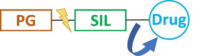
Schematic illustration of the concept of self-immolative linkers. PG = protecting group.

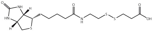
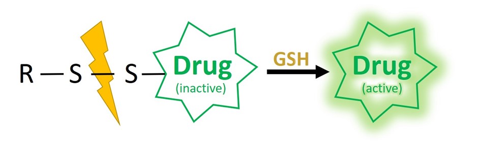
One powerful system relies on disulfide-based self-immolative linkers. The overall red/ox potential in the human blood is oxidative, making disulfide linkages stable during circulation. In contrast, the intracellular milieu of mammalian cells is characterized by an overall reductive potential, thus allowing to revert the disulfide bond formation. Furthermore, cytoplasm typically shows millimolar (1-10 mM) concentrations of the reductant tripeptide glutathione (GSH) in contrast to 2-10 µM in blood and other body fluids.


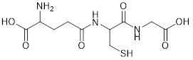
Chemical structure of the tripeptide Glutathione.

Notably, GSH has been recognized as a biomarker of tumor development. Elevated levels are reported inside tumor mass (~100-fold) and cancer cells (~sevenfold) compared to those of normal tissues and cells, respectively. This drastic discrepancy in GSH concentration renders it possible to develop GSH-responsive materials which can be selectively activated in pathological tissues. Those specifications allow for sufficient stability of the linker in the extracellular milieu but spontaneous self-immolative reaction within the cytosol upon GSH-mediated disulfide cleavage.

SILs can be mainly classified into two groups depending on the self-immolative fragmentation process either based on the quinone methide cascade (e.g. 1,6-elimination) or an intramolecular cyclization reaction. In addition, the linker’s chemical composition (e.g. disulfide ethoxycarbonyl (SSE) vs. disulfide benzyloxycarbonyl (SSB)) results in chemically tunable kinetics of the self-immolative cleavage due to different response rate towards GSH, showing higher rates for SSB-based DSILs compared to SSE-based ones. Thus, the choice of the linker allows for fine-tuning of the cleavage speed and payload release.


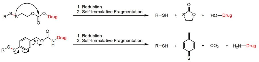
Disulfide-based self-immolative linkers either based on an intramolecular cyclization cascade or an 1,6-elimination.

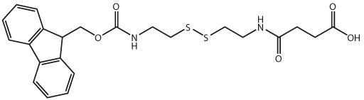
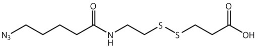
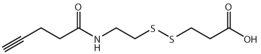
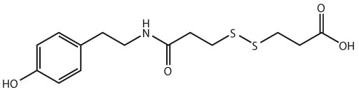
Please find many different disulfide-based self-immolative linkers within our section Related Products (see below). If you cannot find your desired derivative, please contact us for a custom synthesis!

Interested in further information on linker technologies? -> Please feel free to download our new brochure on Linkerology!


References:

  • Self-immolative Linkers Literally Bridge Disulfide Chemistry and the Realm of Thiol-Free Drugs; C. F. Riber; A. A. A. Smith, A. N. Zelikin; Adv Healthcare Mater 2015; 4(12): 1887-1890. https://doi.org/10.1002/adhm.201500344.
  • Labeling Carboxyl Groups of Surface-Exposed Proteins Provides an Orthogonal Approach for Cell Surface Isolation; N. E. Ozkan Kucuk, E. Sanal, E. Tan, T. Mitchison, N. Ozlu; J Proteome Res 2018; 17: 1784-1793. https://doi.org/10.1021/acs.jproteome.7b00825.
  • Development of trifunctional probes for glycoproteomic analysis; C. S. Tsai, P. Y. Liu, H. Y. Yen, T. L. Hsu, C. H. Wong; Chem. Commun. 2010; 46: 5575-7. https://doi.org/10.1039/c0cc00345j.
  • Fluorometric assay for tissue transglutaminase-mediated transamidation activity; C. Gnaccarini, W. Ben-Tahar, W. D. Lubell, J. N. Pelletier, J. W. Keillor; Bioorg Med Chem 2009; 17: 6354-9. https://doi.org/10.1016/j.bmc.2009.07.031.
  • Convenient synthesis of photoaffinity probes and evaluation of their labeling abilities; T. Kan, Y. Kita, Y. Morohashi, Y. Tominari, S. Hosoda, T. Tomita, H. Natsugari, T. Iwatsubo, T. Fukuyama; Org Lett. 2007; 9: 2055-8. https://doi.org/10.1021/ol070376i.
  • Facile synthesis toward the construction of an activity probe library for glycosidases; T. H. Shie, Y. L. Chiang, J. J. Lin, Y. K. Li, L. C. Lo; Carbohydr Res 2006; 341: 443-56. https://doi.org/10.1016/j.carres.2005.12.005.
  • A simple photo-affinity labeling protocol; H.-Y. Li, Y. Liu, K. Fang, K. Nakanishi; Chem. Comm. 1999; 365-366. https://doi.org/10.1039/a809507h.
  • Linker Technologies for Antibody-Drug Conjugates; B. Nolting; Antibody-Drug Conjugates L. Ducry 2013; 1045: 71-100. https://doi.org/10.1007/978-1-62703-541-5_5.
  • Disulfide-Based Self-Immolative Linkers and Functional Bioconjugates for Biological Applications; Z. Deng, J. Hu, S. Liu; Macromol Rapid Commun 2020; 41: e1900531. https://doi.org/10.1002/marc.201900531.
  • Reduction-Triggered Transformation of Disulfide-Containing Micelles at Chemically Tunable Rates; Z. Deng, S. Yuan, R. X. Xu, H. Liang, S. Liu; Angew. Chem. Int. Ed. 2018; 57: 8896-8900. https://doi.org/10.1002/anie.201802909.
  • Modulated Fragmentation of Proapoptotic Peptide Nanoparticles Regulates Cytotoxicity; T. Suma, J. Cui, M. Müllner, S. Fu, J. Tran, K. F. Noi, Y. Ju, F. Caruso; J. Am. Chem. Soc. 2017; 139: 4009-4018. https://doi.org/10.1021/jacs.6b1302.
  • Engineering Intracellular Delivery Nanocarriers and Nanoreactors from Oxidation-Responsive Polymersomes via Synchronized Bilayer Cross-Linking and Permeabilizing Inside Live Cells; Z. Deng, Y. Qian, Y. Yu, G. Liu, J. Hu, G. Zhang, S. Liu; J. Am. Chem. Soc. 2016; 138: 10452-10466. https://doi.org/10.1021/jacs.6b04115.
Related Products
    1. Structure image for Boc-Cystamine*HCl
      Boc-Cystamine*HCl

      Product code: BNN1063

      Starting at €90.00

    2. Structure image for Boc-Cystamine-Suc-OH
      Boc-Cystamine-Suc-OH

      Product code: BAA2180

      Starting at €145.00

    3. Structure image for N3-Cystamine-Suc-OSu
      N3-Cystamine-Suc-OSu

      Product code: HAA2255

      Starting at €171.00

    4. Structure image for Boc-Cystamine
      Boc-Cystamine

      Product code: BNN1170

      Starting at €81.00

    5. Structure image for Biotin-SS-Tyramide
      Biotin-SS-Tyramide

      Product code: LS-3570

      Starting at €350.00

    6. Structure image for Boc-SS-COOH
      Boc-SS-COOH

      Product code: RL-2190

      Starting at €200.00

    7. Structure image for Fmoc-SS-COOH
      Fmoc-SS-COOH

      Product code: RL-2200

      Starting at €625.00

    8. Structure image for N3-Cystamine*HCl
      N3-Cystamine*HCl

      Product code: HNN1090

      Starting at €90.00

    9. Structure image for Fmoc-AEDI-OH
      Fmoc-AEDI-OH

      Product code: RL-2800

      Starting at €720.00

    10. Structure image for Boc-AEDI-OH
      Boc-AEDI-OH

      Product code: RL-2810

      Starting at €470.00

    11. Structure image for Biotin-PEG(4)-SS-Tyramide
      Biotin-PEG(4)-SS-Tyramide

      Product code: LS-3930

      Starting at €350.00

    12. Biotin-SS-COOH
      Biotin-SS-COOH

      Product code: RL-3300.0250

      €325.00
    13. Biotin-SS-COOH
      Biotin-SS-COOH

      Product code: RL-3300

      Starting at €325.00

    14. Biotin-PEG(4)-SS-COOH
      Biotin-PEG(4)-SS-COOH

      Product code: PEG8090

      Starting at €100.00

    15. Structure image for Biotin-PEG(4)-SS-Azide
      Biotin-PEG(4)-SS-Azide

      Product code: PEG8100

      Starting at €225.00

    16. Biotin-PEG(4)-SS-Alkyne
      Biotin-PEG(4)-SS-Alkyne

      Product code: PEG8110

      Starting at €350.00

    17. Biotin-PEG(4)-SS-DBCO
      Biotin-PEG(4)-SS-DBCO

      Product code: PEG8120

      Starting at €225.00

    18. Fmoc-Cystamine-Suc
      Fmoc-Cystamine-Suc

      Product code: RL-3310

      Starting at €99.00

    19. Azido-Pen-SS-COOH
      Azido-Pen-SS-COOH

      Product code: RL-3320

      Starting at €325.00

    20. Alkyne-SS-COOH
      Alkyne-SS-COOH

      Product code: RL-3330

      Starting at €325.00

    21. Tyramide-SS-COOH
      Tyramide-SS-COOH

      Product code: LS-4010

      Starting at Please inquire

    22. Tyramide-SS-amine*HCl
      Tyramide-SS-amine*HCl

      Product code: LS-3960

      Starting at €245.00

    23. Structure image for Fmoc-Cystamine*HCl
      Fmoc-Cystamine*HCl

      Product code: RL-3370

      Starting at €126.00

    24. Structure image for Biotin-cystamine*TFA
      Biotin-cystamine*TFA

      Product code: RL-3391

      Starting at €145.00

    25. Structure image for Boc-NH-SS-OH
      Boc-NH-SS-OH

      Product code: RL-3510

      Starting at €275.00

    26. Structure image for Boc-NH-SS-OpNC
      Boc-NH-SS-OpNC

      Product code: RL-3520

      Starting at €175.00

    27. Structure image for Fmoc-NH-SS-OH
      Fmoc-NH-SS-OH

      Product code: RL-3530

      Starting at €275.00

    28. Structure image for Fmoc-NH-SS-OpNC
      Fmoc-NH-SS-OpNC

      Product code: RL-3540

      Starting at €175.00

    29. Structure image for Boc-NH-SS-Bzl-OH
      Boc-NH-SS-Bzl-OH

      Product code: RL-3560

      Starting at Please inquire

    30. Structure image for Boc-NH-SS-Bzl-OpNC
      Boc-NH-SS-Bzl-OpNC

      Product code: RL-3570

      Starting at Please inquire

    31. Structure image for Fmoc-NH-SS-Bzl-OH
      Fmoc-NH-SS-Bzl-OH

      Product code: RL-3580

      Starting at €250.00